Half Value layer (HVL)
About[]
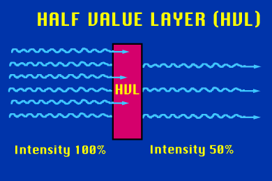
Half Value Layer (HVL) or beam quality is the thickness of any given material where 50% of the incident energy has been attenuated is know as the half-value layer (HVL). The HVL is expressed in units of distance (mm or cm). Like the attenuation coefficient, it is photon energy dependent. Increasing the penetrating energy of a stream of photons will result in an increase in a material's HVL.
The HVL is inversely proportional to the attenuation coefficient. If an incident energy of 1 and a transmitted energy is 0.5 is plugged into the equation introduced on the preceding page, it can be seen that the HVL multiplied by m must equal 0.693.[1]
Formula:
- I = Ioe-μx
where Io is the incident intensity, that is, I = Io when x = 0. I is the intensity left after the radiation traverses a thickness x of material and e = 2.718, Leonhard Euler started to use the letter e for the constant in 1727 or 1728,[5] and the first use of e in a publication was Euler's Mechanica (1736), is the base of the system of natural logarithms.
Shielding thickness[]
The thickness of lead required to achieve the same shielding effect against radiation, under specified conditions, as that provided by a given material.
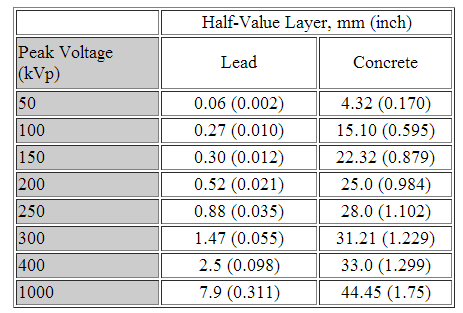
Approximate Half-Value Layer for Various Materials when Radiation is from an X-ray Source (e.g. xray tube materiel such as tungsten (W), Molybdenum (Mo),etc. Great estimation for room shielding thickness preventing population exposure to scatter radiation.
FDA Requirements[]
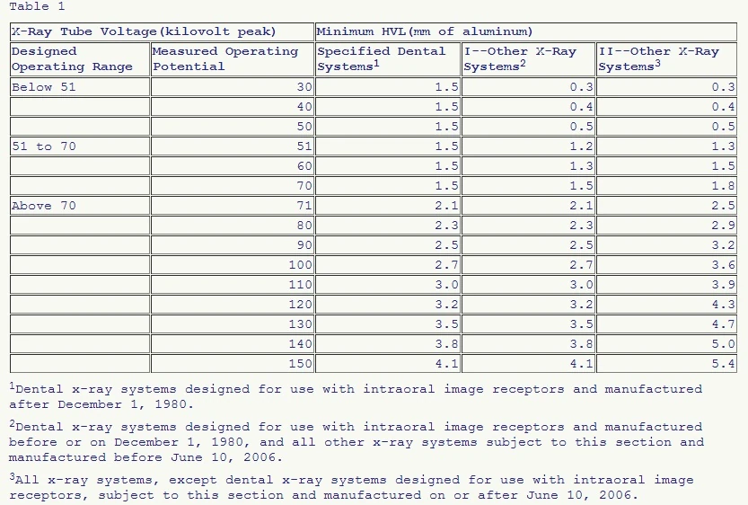
Half-value layer (HVL) or beam quality . The HVL of the useful beam for a given x-ray tube potential shall not be less than the appropriate value shown in table 1 in paragraph (m)(1) of this section under the heading "Specified Dental Systems," for any dental x-ray system designed for use with intraoral image receptors and manufactured after December 1, 1980; under the heading "I--Other X-Ray Systems," for any dental x-ray system designed for use with intraoral image receptors and manufactured before December 1, 1980, and all other x-ray systems subject to this section and manufactured before June 10, 2006; and under the heading "II--Other X-Ray Systems," for all x-ray systems, except dental x-ray systems designed for use with intraoral image receptors, subject to this section and manufactured on or after June 10, 2006.
Half-value layer requirement chart from 21 CFR Sub-chapter J, Title 21, Volume 8]. [Revised as of April 1, 2013]
If it is necessary to determine such HVL at an x-ray tube potential which is not listed in table 1 in paragraph (m)(1) of this section, linear interpolation or extrapolation may be made. Positive means shall be provided to ensure that at least the minimum filtration needed to achieve the above beam quality requirements is in the useful beam during each exposure.[2] In order to measure HVL, the corresponding x-ray tube voltages (kVp)s must match the minimum aluminin requirments to achive a 50% half life measurement. For example, lets image we are measureeing HVL and have our x-ray techniques set to 90kVp, 100mA at 1 second timing. Using the following table from 21 CFR 70kVp is the above the column called "above 70". Next, we look at the 70kVp in the "measured operating potential"column which is 71 and then scroll to the 3rd column on the far right called "II-Other xray systems" and the most amount of aluminum we can use is 2.5mmAL for our final measurement.

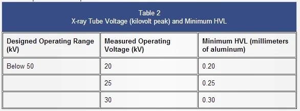
Half Value Layer (HVL)requirements for mammography units
The HVL shall meet the specifications of Sec. 1020.30(m)(1) for ammorgrapght devices for the minimum HVL.
Calculations[]
Traditionally, half‐value layer (HVL) of an x‐ray beam has been calculated through either linear (Eq. 2) or semi‐logarithmic (Eq. 3) interpolation under the assumption that attenuation varies linearly or monochromatically over a small range of attenuation thicknesses. Our aim is to evaluate the accuracy of HVL calculation using Lambert W interpolation. [3]
References[]
- ↑ NDT Resource Center. Half Value layer. http://www.ndt-ed.org/EducationResources/CommunityCollege/Radiography/Physics/HalfValueLayer.htm
- ↑ TITLE 21--FOOD AND DRUGS CHAPTER I--FOOD AND DRUG ADMINISTRATION. DEPARTMENT OF HEALTH AND HUMAN SERVICES SUBCHAPTER J--RADIOLOGICAL HEALTH PART 1020 -- PERFORMANCE STANDARDS FOR IONIZING RADIATION EMITTING PRODUCTS. Revised as of April 1, 2013. http://www.accessdata.fda.gov/scripts/cdrh/cfdocs/cfcfr/CFRSearch.cfm?FR=1020.30
- ↑ K Boitnott, S Kappadath, A White, N Atkinson, and D Cody. A New Method for Calculating the Half‐Value Layer of X‐Ray Beams. Medical Physics. http://online.medphys.org/resource/1/mphya6/v36/i6/p2471_s3?isAuthorized=no